South Florida MRI Centers
South Florida MRI Centers
South Florida
MRI Centers
Quick precision imaging at patient friendly ACR accredited facilities in Broward and Miami-Dade.
Quick precision imaging at patient friendly ACR accredited facilities in Broward and Miami-Dade.
Quick precision imaging at patient friendly ACR accredited facilities in Broward and Miami-Dade.
Schedule your
MRI appointment
We'll confirm your appointment within 2 hours. For urgent matters, please call or text us directly at
+1 (305) 654-7725
+1 (954) 792-0777
Schedule your
MRI appointment
We'll confirm your appointment within 2 hours. For urgent matters, please call or text us directly at
+1 (305) 654-7725
+1 (954) 792-0777
Schedule your
MRI appointment
We'll confirm your appointment within 2 hours. For urgent matters, please call or text us directly at
+1 (305) 654-7725
+1 (954) 792-0777
Professional & Compassionate
every step of the way
Accu-Med Diagnostics is committed to delivering the highest standard of professional medical imaging to patients, referring physicians, and healthcare provider systems alike.

0+
0+
Years of experience
0+
0+
Years of experience
0+
0+
Years of experience
9,000+
9,000+
Happy patients
9,000+
9,000+
Happy patients
9,000+
9,000+
Happy patients
Common Appointment Types and Times
01
Cervical (Neck) MRI
Starting at 20 Minutes
A neck MRI is a powerful diagnostic tool used to evaluate the soft tissues, spinal structures, and blood vessels in the cervical spine. It is commonly recommended for individuals experiencing persistent neck pain, numbness or tingling in the arms, weakness, or after trauma such as whiplash. It also helps detect conditions like herniated discs, spinal stenosis, degenerative disc disease, tumors, and infections. Common findings may include disc bulges, nerve compression, arthritis, spinal cord abnormalities, or soft tissue masses—allowing for accurate diagnosis and effective treatment planning.

02
Thoracic (Mid-Back) MRI
Starting at 25 Minutes
A thoracic MRI is typically ordered to evaluate unexplained mid-back pain, neurological symptoms, or suspected spinal conditions affecting the upper spine. Doctors may recommend this scan for patients experiencing numbness, weakness, coordination issues, or symptoms unresponsive to other treatments. Common findings on a thoracic MRI include herniated or bulging discs, spinal stenosis, degenerative disc disease, spinal cord compression, and myelopathy. It can also detect more serious conditions like tumors, spinal cord cysts (syringomyelia), infections, fractures, or inflammatory diseases. This imaging is essential for accurately diagnosing the cause of symptoms and guiding effective treatment plans.
03
Lumbar (Low Back) MRI
Starting at 20 Minutes
A lumbar MRI is a valuable diagnostic tool used to evaluate lower back pain, sciatica, numbness, weakness, or symptoms following an injury. It’s commonly recommended for patients with chronic back pain, suspected disc herniations, degenerative disc disease, spinal stenosis, or nerve compression. This non-invasive scan provides detailed images of the spine’s bones, discs, nerves, and soft tissues, helping detect common issues such as bulging or herniated discs, arthritis, nerve impingement, spinal canal narrowing, spondylolisthesis, tumors, infections, or fractures. A lumbar MRI plays a key role in diagnosing the cause of symptoms and guiding effective treatment plans.
04
Shoulder MRI
Starting at 30 Minutes
A shoulder MRI is commonly ordered to evaluate pain, stiffness, weakness, or limited range of motion that doesn’t improve with rest or physical therapy. It’s especially useful after injuries, such as rotator cuff tears, labral tears, or dislocations, and helps diagnose chronic conditions like arthritis or tendonitis. This detailed imaging reveals common findings such as rotator cuff tears, labrum injuries, bursitis, tendon inflammation or degeneration, joint effusion, and arthritis. A shoulder MRI is a crucial tool for identifying soft tissue damage, guiding treatment decisions, and helping patients regain strength and mobility.
05
Knee MRI
Starting at 30 Minutes
A knee MRI is commonly performed to diagnose the cause of pain, swelling, instability, or limited movement in the knee joint. It is especially useful after injuries from sports, falls, or accidents, and helps identify conditions like ACL tears, meniscus injuries, ligament sprains, or cartilage damage. It also detects chronic issues such as osteoarthritis, tendonitis, bursitis, joint effusion, and Baker’s cysts. A knee MRI provides detailed images of the bones, ligaments, tendons, cartilage, and soft tissues, making it an essential tool for guiding treatment—whether that’s physical therapy, injections, or surgery.
06
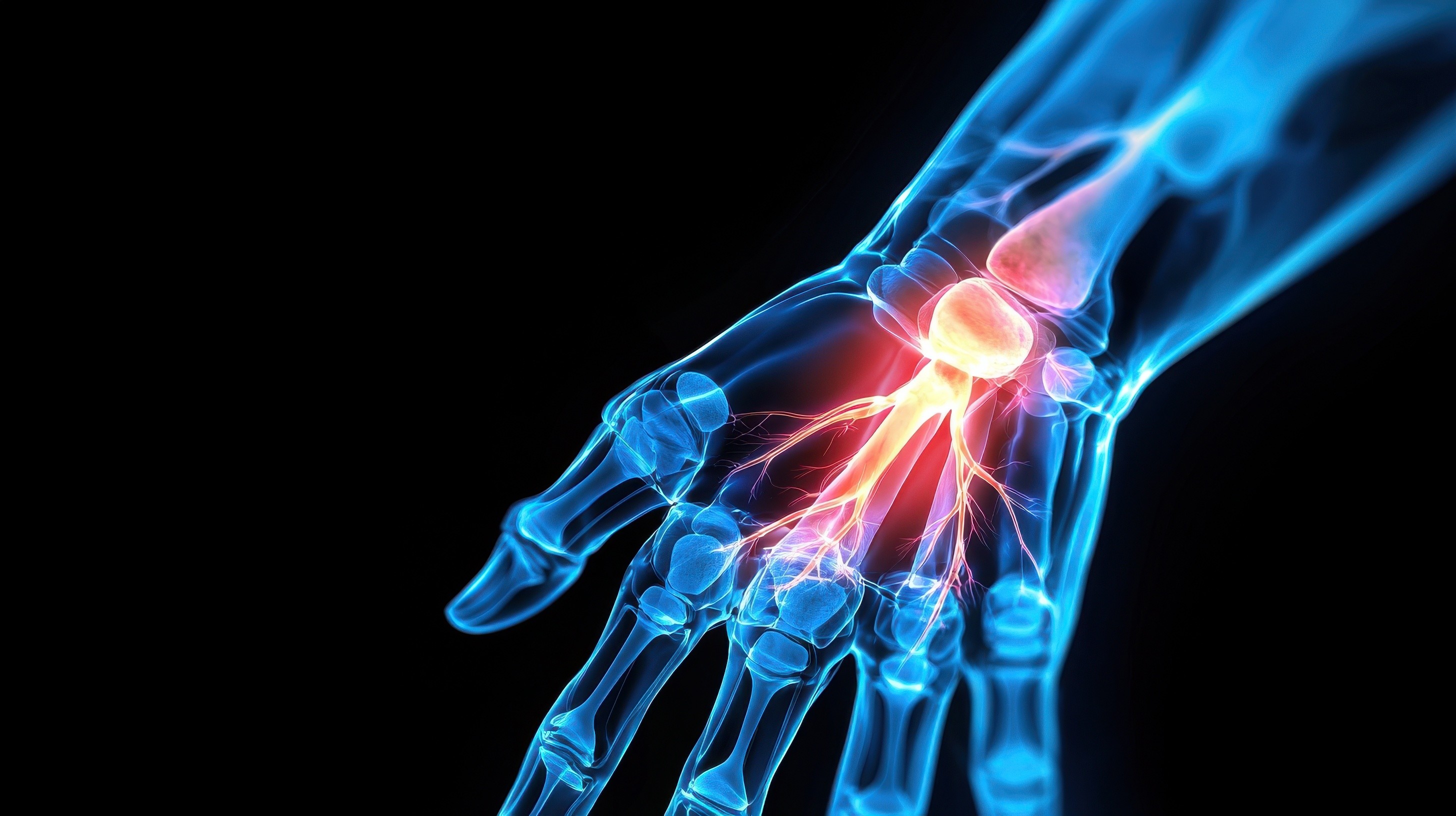
Wrist MRI
Starting at 30 Minutes
A wrist MRI is a non-invasive imaging test that uses powerful magnets and radio waves to create detailed images of the bones, ligaments, tendons, cartilage, and soft tissues in the wrist. It is commonly used to evaluate unexplained wrist pain, injuries from falls, sports-related trauma, or symptoms like swelling, numbness, or weakness. A wrist MRI helps diagnose conditions such as ligament tears, TFCC injuries, tendonitis, carpal tunnel syndrome, arthritis, occult fractures, and ganglion cysts. This advanced imaging tool provides critical information for accurate diagnosis and effective treatment planning, especially when X-rays or physical exams are inconclusive.
07
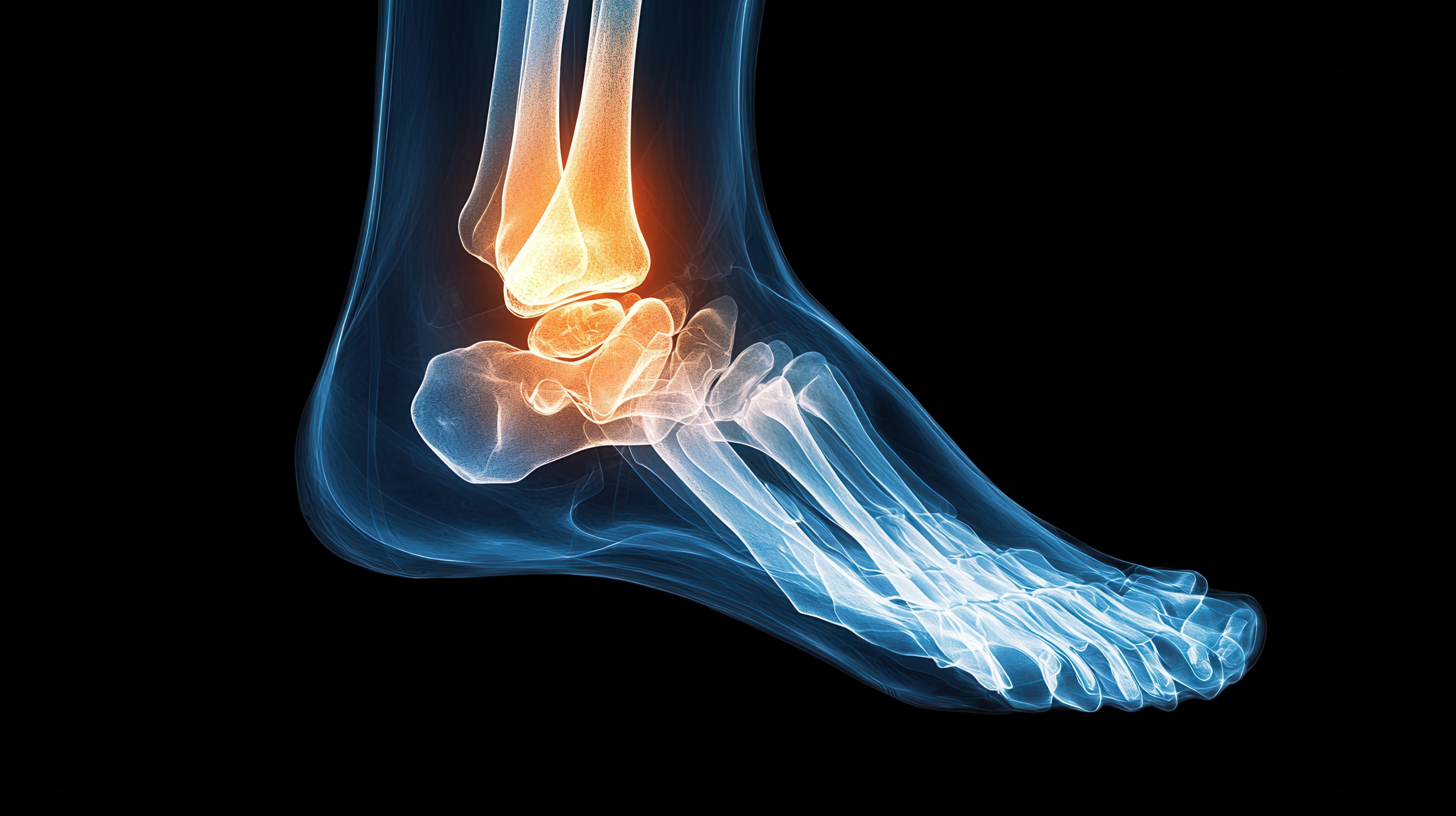
Ankle MRI
Starting at 30 Minutes
An ankle MRI is a non-invasive imaging test that uses powerful magnets and radio waves to produce detailed images of the bones, ligaments, tendons, cartilage, and soft tissues in the ankle. It is commonly used to diagnose the cause of persistent pain, swelling, instability, or injury when X-rays or physical exams are inconclusive. An ankle MRI can detect ligament tears, Achilles tendon injuries, stress fractures, tendonitis, arthritis, and cartilage damage, providing critical information for accurate diagnosis and effective treatment planning.
08
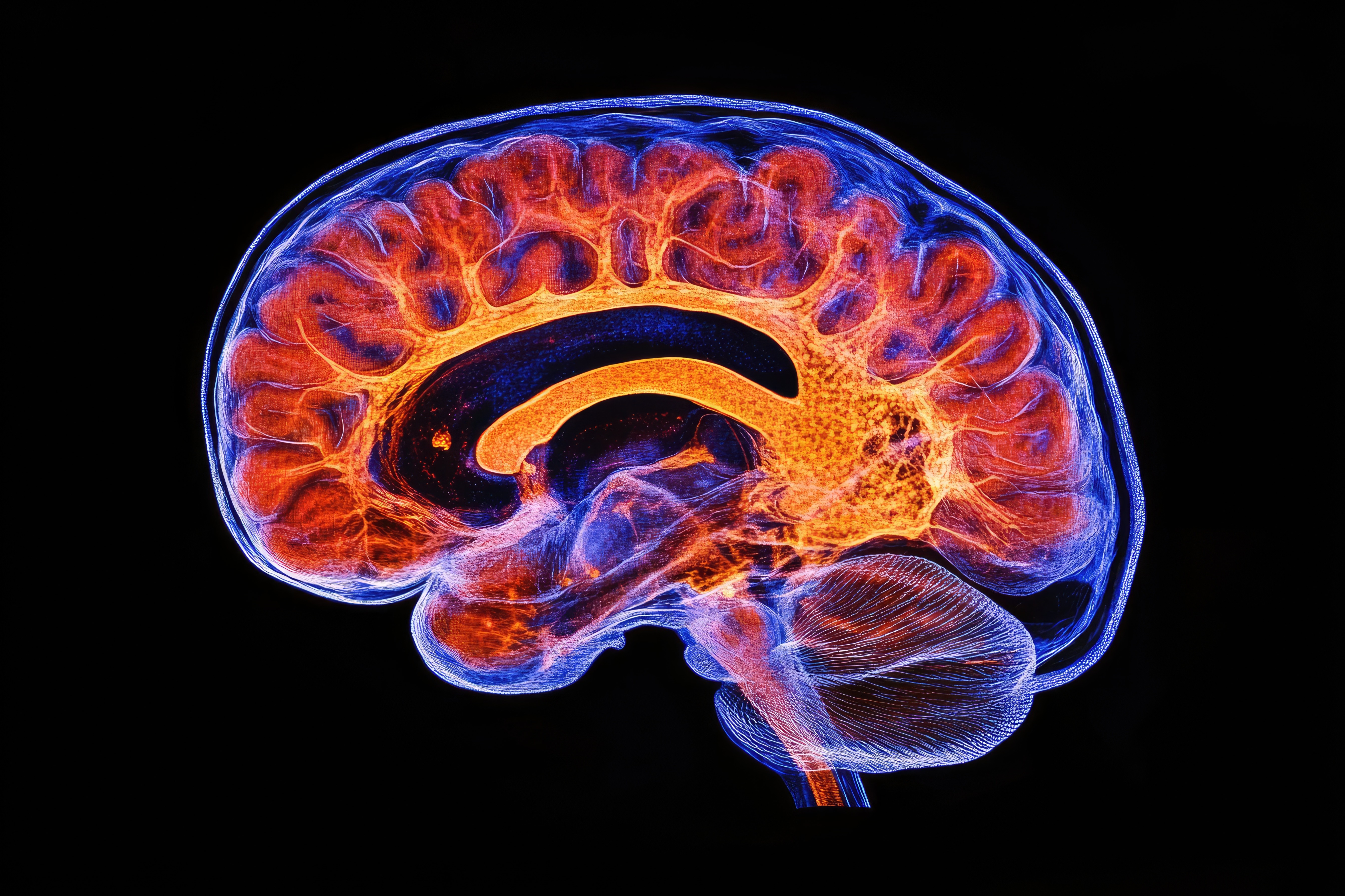
Brain MRI
Starting at 30 Minutes
A brain MRI (Magnetic Resonance Imaging) is a non-invasive imaging test that uses powerful magnets and radio waves to create detailed images of the brain and surrounding structures. Unlike X-rays or CT scans, it does not use radiation and is especially effective for visualizing soft tissues. A brain MRI is commonly used to evaluate symptoms such as headaches, seizures, dizziness, memory problems, or changes in vision and behavior. It helps detect a wide range of conditions, including brain tumors, stroke, multiple sclerosis, infections, aneurysms, brain injuries, and structural abnormalities. This advanced imaging technique provides critical information for accurate diagnosis, monitoring, and treatment planning.
09
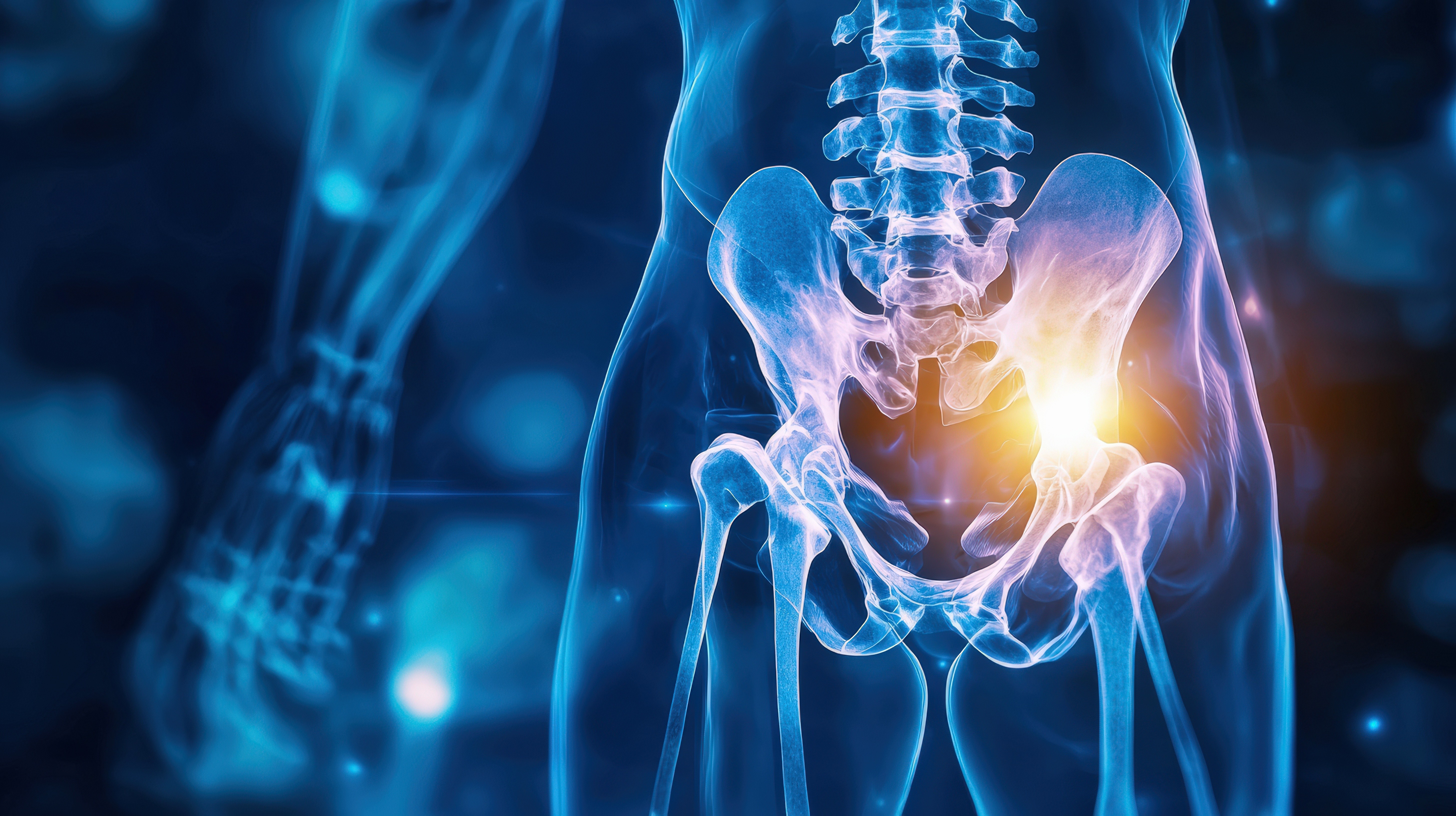
Hip MRI
Starting at 30 Minutes
A hip MRI (Magnetic Resonance Imaging of the hip) is a non-invasive diagnostic test that uses powerful magnets and radio waves to produce detailed images of the bones, cartilage, muscles, tendons, ligaments, and soft tissues surrounding the hip joint. It is commonly used to evaluate persistent hip pain, stiffness, swelling, or limited mobility—especially when X-rays or physical exams do not provide enough information. A hip MRI can detect conditions such as labral tears, hip impingement (FAI), bursitis, tendonitis, stress fractures, arthritis, and avascular necrosis. This advanced imaging tool helps physicians diagnose the root cause of symptoms and develop accurate, targeted treatment plans.
01
Cervical (Neck) MRI
Starting at 20 Minutes
A neck MRI is a powerful diagnostic tool used to evaluate the soft tissues, spinal structures, and blood vessels in the cervical spine. It is commonly recommended for individuals experiencing persistent neck pain, numbness or tingling in the arms, weakness, or after trauma such as whiplash. It also helps detect conditions like herniated discs, spinal stenosis, degenerative disc disease, tumors, and infections. Common findings may include disc bulges, nerve compression, arthritis, spinal cord abnormalities, or soft tissue masses—allowing for accurate diagnosis and effective treatment planning.
02
Thoracic (Mid-Back) MRI
Starting at 25 Minutes
A thoracic MRI is typically ordered to evaluate unexplained mid-back pain, neurological symptoms, or suspected spinal conditions affecting the upper spine. Doctors may recommend this scan for patients experiencing numbness, weakness, coordination issues, or symptoms unresponsive to other treatments. Common findings on a thoracic MRI include herniated or bulging discs, spinal stenosis, degenerative disc disease, spinal cord compression, and myelopathy. It can also detect more serious conditions like tumors, spinal cord cysts (syringomyelia), infections, fractures, or inflammatory diseases. This imaging is essential for accurately diagnosing the cause of symptoms and guiding effective treatment plans.
03
Lumbar (Low Back) MRI
Starting at 20 Minutes
A lumbar MRI is a valuable diagnostic tool used to evaluate lower back pain, sciatica, numbness, weakness, or symptoms following an injury. It’s commonly recommended for patients with chronic back pain, suspected disc herniations, degenerative disc disease, spinal stenosis, or nerve compression. This non-invasive scan provides detailed images of the spine’s bones, discs, nerves, and soft tissues, helping detect common issues such as bulging or herniated discs, arthritis, nerve impingement, spinal canal narrowing, spondylolisthesis, tumors, infections, or fractures. A lumbar MRI plays a key role in diagnosing the cause of symptoms and guiding effective treatment plans.
04
Shoulder MRI
Starting at 30 Minutes
A shoulder MRI is commonly ordered to evaluate pain, stiffness, weakness, or limited range of motion that doesn’t improve with rest or physical therapy. It’s especially useful after injuries, such as rotator cuff tears, labral tears, or dislocations, and helps diagnose chronic conditions like arthritis or tendonitis. This detailed imaging reveals common findings such as rotator cuff tears, labrum injuries, bursitis, tendon inflammation or degeneration, joint effusion, and arthritis. A shoulder MRI is a crucial tool for identifying soft tissue damage, guiding treatment decisions, and helping patients regain strength and mobility.
05
Knee MRI
Starting at 30 Minutes
A knee MRI is commonly performed to diagnose the cause of pain, swelling, instability, or limited movement in the knee joint. It is especially useful after injuries from sports, falls, or accidents, and helps identify conditions like ACL tears, meniscus injuries, ligament sprains, or cartilage damage. It also detects chronic issues such as osteoarthritis, tendonitis, bursitis, joint effusion, and Baker’s cysts. A knee MRI provides detailed images of the bones, ligaments, tendons, cartilage, and soft tissues, making it an essential tool for guiding treatment—whether that’s physical therapy, injections, or surgery.
06
Wrist MRI
Starting at 30 Minutes
A wrist MRI is a non-invasive imaging test that uses powerful magnets and radio waves to create detailed images of the bones, ligaments, tendons, cartilage, and soft tissues in the wrist. It is commonly used to evaluate unexplained wrist pain, injuries from falls, sports-related trauma, or symptoms like swelling, numbness, or weakness. A wrist MRI helps diagnose conditions such as ligament tears, TFCC injuries, tendonitis, carpal tunnel syndrome, arthritis, occult fractures, and ganglion cysts. This advanced imaging tool provides critical information for accurate diagnosis and effective treatment planning, especially when X-rays or physical exams are inconclusive.
07
Ankle MRI
Starting at 30 Minutes
An ankle MRI is a non-invasive imaging test that uses powerful magnets and radio waves to produce detailed images of the bones, ligaments, tendons, cartilage, and soft tissues in the ankle. It is commonly used to diagnose the cause of persistent pain, swelling, instability, or injury when X-rays or physical exams are inconclusive. An ankle MRI can detect ligament tears, Achilles tendon injuries, stress fractures, tendonitis, arthritis, and cartilage damage, providing critical information for accurate diagnosis and effective treatment planning.
08
Brain MRI
Starting at 30 Minutes
A brain MRI (Magnetic Resonance Imaging) is a non-invasive imaging test that uses powerful magnets and radio waves to create detailed images of the brain and surrounding structures. Unlike X-rays or CT scans, it does not use radiation and is especially effective for visualizing soft tissues. A brain MRI is commonly used to evaluate symptoms such as headaches, seizures, dizziness, memory problems, or changes in vision and behavior. It helps detect a wide range of conditions, including brain tumors, stroke, multiple sclerosis, infections, aneurysms, brain injuries, and structural abnormalities. This advanced imaging technique provides critical information for accurate diagnosis, monitoring, and treatment planning.
09
Hip MRI
Starting at 30 Minutes
A hip MRI (Magnetic Resonance Imaging of the hip) is a non-invasive diagnostic test that uses powerful magnets and radio waves to produce detailed images of the bones, cartilage, muscles, tendons, ligaments, and soft tissues surrounding the hip joint. It is commonly used to evaluate persistent hip pain, stiffness, swelling, or limited mobility—especially when X-rays or physical exams do not provide enough information. A hip MRI can detect conditions such as labral tears, hip impingement (FAI), bursitis, tendonitis, stress fractures, arthritis, and avascular necrosis. This advanced imaging tool helps physicians diagnose the root cause of symptoms and develop accurate, targeted treatment plans.
01
Cervical (Neck) MRI
A neck MRI is a powerful diagnostic tool used to evaluate the soft tissues, spinal structures, and blood vessels in the cervical spine. It is commonly recommended for individuals experiencing persistent neck pain, numbness or tingling in the arms, weakness, or after trauma such as whiplash. It also helps detect conditions like herniated discs, spinal stenosis, degenerative disc disease, tumors, and infections. Common findings may include disc bulges, nerve compression, arthritis, spinal cord abnormalities, or soft tissue masses—allowing for accurate diagnosis and effective treatment planning.
02
Thoracic (Mid-Back) MRI
A thoracic MRI is typically ordered to evaluate unexplained mid-back pain, neurological symptoms, or suspected spinal conditions affecting the upper spine. Doctors may recommend this scan for patients experiencing numbness, weakness, coordination issues, or symptoms unresponsive to other treatments. Common findings on a thoracic MRI include herniated or bulging discs, spinal stenosis, degenerative disc disease, spinal cord compression, and myelopathy. It can also detect more serious conditions like tumors, spinal cord cysts (syringomyelia), infections, fractures, or inflammatory diseases. This imaging is essential for accurately diagnosing the cause of symptoms and guiding effective treatment plans.
03
Lumbar (Low Back) MRI
A lumbar MRI is a valuable diagnostic tool used to evaluate lower back pain, sciatica, numbness, weakness, or symptoms following an injury. It’s commonly recommended for patients with chronic back pain, suspected disc herniations, degenerative disc disease, spinal stenosis, or nerve compression. This non-invasive scan provides detailed images of the spine’s bones, discs, nerves, and soft tissues, helping detect common issues such as bulging or herniated discs, arthritis, nerve impingement, spinal canal narrowing, spondylolisthesis, tumors, infections, or fractures. A lumbar MRI plays a key role in diagnosing the cause of symptoms and guiding effective treatment plans.
04
Shoulder MRI
A shoulder MRI is commonly ordered to evaluate pain, stiffness, weakness, or limited range of motion that doesn’t improve with rest or physical therapy. It’s especially useful after injuries, such as rotator cuff tears, labral tears, or dislocations, and helps diagnose chronic conditions like arthritis or tendonitis. This detailed imaging reveals common findings such as rotator cuff tears, labrum injuries, bursitis, tendon inflammation or degeneration, joint effusion, and arthritis. A shoulder MRI is a crucial tool for identifying soft tissue damage, guiding treatment decisions, and helping patients regain strength and mobility.
05
Knee MRI
A knee MRI is commonly performed to diagnose the cause of pain, swelling, instability, or limited movement in the knee joint. It is especially useful after injuries from sports, falls, or accidents, and helps identify conditions like ACL tears, meniscus injuries, ligament sprains, or cartilage damage. It also detects chronic issues such as osteoarthritis, tendonitis, bursitis, joint effusion, and Baker’s cysts. A knee MRI provides detailed images of the bones, ligaments, tendons, cartilage, and soft tissues, making it an essential tool for guiding treatment—whether that’s physical therapy, injections, or surgery.
06
Wrist MRI
A wrist MRI is a non-invasive imaging test that uses powerful magnets and radio waves to create detailed images of the bones, ligaments, tendons, cartilage, and soft tissues in the wrist. It is commonly used to evaluate unexplained wrist pain, injuries from falls, sports-related trauma, or symptoms like swelling, numbness, or weakness. A wrist MRI helps diagnose conditions such as ligament tears, TFCC injuries, tendonitis, carpal tunnel syndrome, arthritis, occult fractures, and ganglion cysts. This advanced imaging tool provides critical information for accurate diagnosis and effective treatment planning, especially when X-rays or physical exams are inconclusive.
07
Ankle MRI
An ankle MRI is a non-invasive imaging test that uses powerful magnets and radio waves to produce detailed images of the bones, ligaments, tendons, cartilage, and soft tissues in the ankle. It is commonly used to diagnose the cause of persistent pain, swelling, instability, or injury when X-rays or physical exams are inconclusive. An ankle MRI can detect ligament tears, Achilles tendon injuries, stress fractures, tendonitis, arthritis, and cartilage damage, providing critical information for accurate diagnosis and effective treatment planning.
08
Brain MRI
A brain MRI (Magnetic Resonance Imaging) is a non-invasive imaging test that uses powerful magnets and radio waves to create detailed images of the brain and surrounding structures. Unlike X-rays or CT scans, it does not use radiation and is especially effective for visualizing soft tissues. A brain MRI is commonly used to evaluate symptoms such as headaches, seizures, dizziness, memory problems, or changes in vision and behavior. It helps detect a wide range of conditions, including brain tumors, stroke, multiple sclerosis, infections, aneurysms, brain injuries, and structural abnormalities. This advanced imaging technique provides critical information for accurate diagnosis, monitoring, and treatment planning.
09
Hip MRI
A hip MRI (Magnetic Resonance Imaging of the hip) is a non-invasive diagnostic test that uses powerful magnets and radio waves to produce detailed images of the bones, cartilage, muscles, tendons, ligaments, and soft tissues surrounding the hip joint. It is commonly used to evaluate persistent hip pain, stiffness, swelling, or limited mobility—especially when X-rays or physical exams do not provide enough information. A hip MRI can detect conditions such as labral tears, hip impingement (FAI), bursitis, tendonitis, stress fractures, arthritis, and avascular necrosis. This advanced imaging tool helps physicians diagnose the root cause of symptoms and develop accurate, targeted treatment plans.
What makes us different
Board Certified Techs. ACR Approved Facilities.
Board Certified Techs. ACR Approved Facilities.
Board Certified Techs. ACR Approved Facilities.
Patient Centric Approach to Imaging
Patient Centric Approach to Imaging
Patient Centric Approach to Imaging
Advanced Imaging Technology and Precision Protocols
Advanced Imaging Technology and Precision Protocols
Advanced Imaging Technology and Precision Protocols
Fast Reports, Records & Billing Requests
Fast Reports, Records & Billing Requests
Fast Reports, Records & Billing Requests
Digital Imaging and Wellness Blog
Stay up to date with these useful articles and resources.

Injury
Dec 10, 2025
When the Body Compensates — How One Injury Can Set the Stage for Another
Injury
Dec 10, 2025
When the Body Compensates — How One Injury Can Set the Stage for Another

South Florida
Sep 30, 2025
Accu-Med Diagnostic Centers Weigh in on Tyreek Hill’s Knee Injury and the Vital Role of Imaging in Recovery
South Florida
Sep 30, 2025
Accu-Med Diagnostic Centers Weigh in on Tyreek Hill’s Knee Injury and the Vital Role of Imaging in Recovery

MRI
Sep 15, 2025
What Is an MRI and When Do You Need One?
MRI
Sep 15, 2025
What Is an MRI and When Do You Need One?
Got questions?
We’ve got answers
Everything you need to know about our locations, pricing, and process.
How Long Does it Take?
An MRI scan typically takes between 15 to 60 minutes, depending on the area being examined and whether contrast dye is used.
Does it Hurt? Is it Loud? Is it Cold?
No, MRI scans are painless. You won’t feel the magnetic fields or radio waves. You may hear loud tapping noises during the scan, but earplugs or headphones are usually provided for comfort. The magnet rooms are kept cold, fresh blankets are provided if needed.
How Should I Dress?
Wear loose, comfortable clothing without any metal, such as zippers, buttons, or snaps. You may be asked to change into a gown depending on the area being scanned. Be sure to remove all jewelry, watches, and other metal items before the scan.
What Can You See on a MRI?
MRI scans provide detailed images of soft tissues, organs, bones, and joints. They are especially useful for viewing the brain, spine, muscles, ligaments, tendons, and internal organs. MRI helps diagnose a wide range of conditions, from injuries and inflammation to tumors and neurological disorders.
How Much Does it Cost?
Please call the office for our current self-pay prices. We are not medicare providers.
Can I eat or drink before my MRI?
In most cases, yes. However, if your MRI requires contrast or focuses on your abdomen, you may be asked to avoid eating or drinking a few hours before. We’ll provide specific instructions when you book your appointment.
Is it safe to get an MRI if I have metal in my body?
Please inform us if you have any metal implants, pacemakers, or surgical clips. We will evaluate your situation to ensure your safety.
Will I be exposed to radiation from the MRI scan?
No, MRI scans do not use radiation. Unlike X-rays or CT scans, MRIs use magnetic fields and radio waves to create images, making them a safe option—especially for people who need repeated imaging or are sensitive to radiation.