

Our Diagnostic Services
At Accu-Med Diagnostic Centers, we provide advanced diagnostic imaging services in Miami and Broward to support patients and physicians with fast, accurate results. Our state-of-the-art facilities are equipped with the latest MRI technology, offering a full range of studies including cervical MRI, thoracic MRI, lumbar MRI, brain MRI, and joint MRI. Each exam is performed by experienced technologists and interpreted by board-certified radiologists to ensure the highest level of precision.
We understand that diagnostic testing can feel overwhelming, which is why our team is committed to delivering a comfortable, stress-free experience from start to finish. Whether you’re visiting our Miami MRI center or our Broward MRI center, you’ll receive compassionate care, flexible scheduling, and quick turnaround times on results. At Accu-Med, our mission is simple: to combine cutting-edge imaging with patient-centered service, helping you and your doctor make confident healthcare decisions.
01
Cervical (Neck) MRI
Starting at 20 Minutes
A neck MRI is a powerful diagnostic tool used to evaluate the soft tissues, spinal structures, and blood vessels in the cervical spine. It is commonly recommended for individuals experiencing persistent neck pain, numbness or tingling in the arms, weakness, or after trauma such as whiplash. It also helps detect conditions like herniated discs, spinal stenosis, degenerative disc disease, tumors, and infections. Common findings may include disc bulges, nerve compression, arthritis, spinal cord abnormalities, or soft tissue masses—allowing for accurate diagnosis and effective treatment planning.

02
Thoracic (Mid-Back) MRI
Starting at 25 Minutes
A thoracic MRI is typically ordered to evaluate unexplained mid-back pain, neurological symptoms, or suspected spinal conditions affecting the upper spine. Doctors may recommend this scan for patients experiencing numbness, weakness, coordination issues, or symptoms unresponsive to other treatments. Common findings on a thoracic MRI include herniated or bulging discs, spinal stenosis, degenerative disc disease, spinal cord compression, and myelopathy. It can also detect more serious conditions like tumors, spinal cord cysts (syringomyelia), infections, fractures, or inflammatory diseases. This imaging is essential for accurately diagnosing the cause of symptoms and guiding effective treatment plans.
03
Lumbar (Low Back) MRI
Starting at 20 Minutes
A lumbar MRI is a valuable diagnostic tool used to evaluate lower back pain, sciatica, numbness, weakness, or symptoms following an injury. It’s commonly recommended for patients with chronic back pain, suspected disc herniations, degenerative disc disease, spinal stenosis, or nerve compression. This non-invasive scan provides detailed images of the spine’s bones, discs, nerves, and soft tissues, helping detect common issues such as bulging or herniated discs, arthritis, nerve impingement, spinal canal narrowing, spondylolisthesis, tumors, infections, or fractures. A lumbar MRI plays a key role in diagnosing the cause of symptoms and guiding effective treatment plans.
04
Shoulder MRI
Starting at 30 Minutes
A shoulder MRI is commonly ordered to evaluate pain, stiffness, weakness, or limited range of motion that doesn’t improve with rest or physical therapy. It’s especially useful after injuries, such as rotator cuff tears, labral tears, or dislocations, and helps diagnose chronic conditions like arthritis or tendonitis. This detailed imaging reveals common findings such as rotator cuff tears, labrum injuries, bursitis, tendon inflammation or degeneration, joint effusion, and arthritis. A shoulder MRI is a crucial tool for identifying soft tissue damage, guiding treatment decisions, and helping patients regain strength and mobility.
05
Knee MRI
Starting at 30 Minutes
A knee MRI is commonly performed to diagnose the cause of pain, swelling, instability, or limited movement in the knee joint. It is especially useful after injuries from sports, falls, or accidents, and helps identify conditions like ACL tears, meniscus injuries, ligament sprains, or cartilage damage. It also detects chronic issues such as osteoarthritis, tendonitis, bursitis, joint effusion, and Baker’s cysts. A knee MRI provides detailed images of the bones, ligaments, tendons, cartilage, and soft tissues, making it an essential tool for guiding treatment—whether that’s physical therapy, injections, or surgery.
06
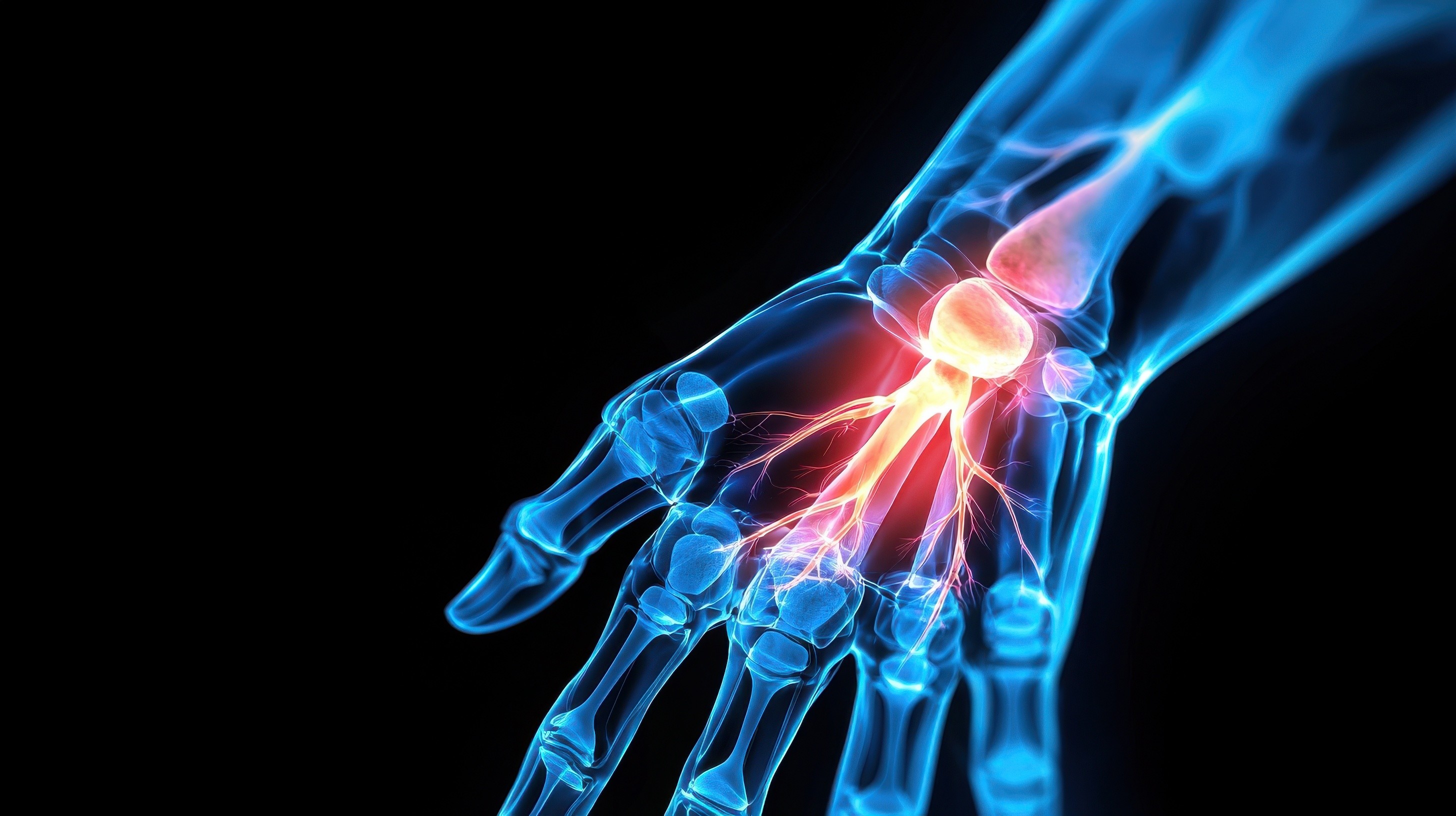
Wrist MRI
Starting at 30 Minutes
A wrist MRI is a non-invasive imaging test that uses powerful magnets and radio waves to create detailed images of the bones, ligaments, tendons, cartilage, and soft tissues in the wrist. It is commonly used to evaluate unexplained wrist pain, injuries from falls, sports-related trauma, or symptoms like swelling, numbness, or weakness. A wrist MRI helps diagnose conditions such as ligament tears, TFCC injuries, tendonitis, carpal tunnel syndrome, arthritis, occult fractures, and ganglion cysts. This advanced imaging tool provides critical information for accurate diagnosis and effective treatment planning, especially when X-rays or physical exams are inconclusive.
07
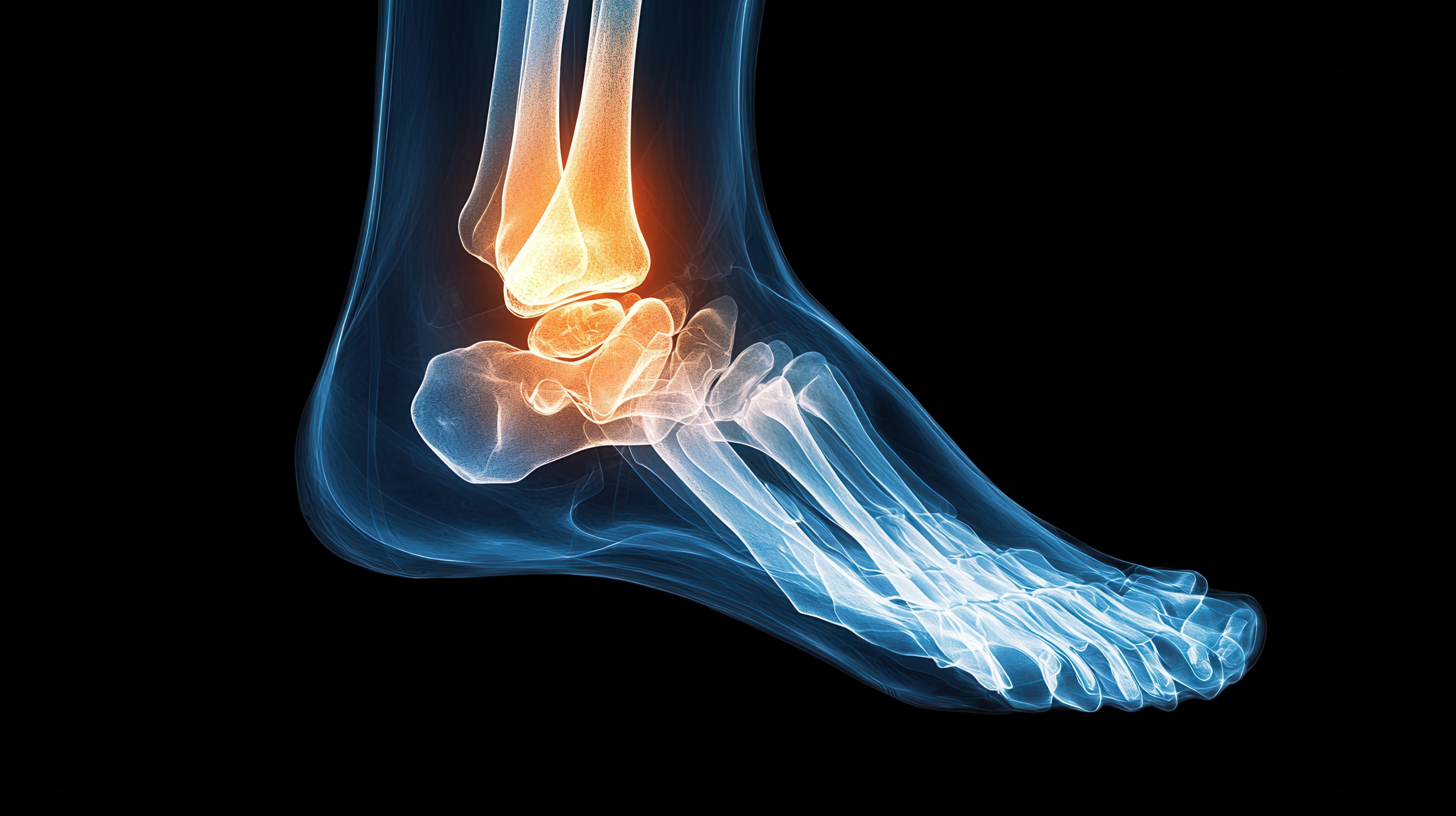
Ankle MRI
Starting at 30 Minutes
An ankle MRI is a non-invasive imaging test that uses powerful magnets and radio waves to produce detailed images of the bones, ligaments, tendons, cartilage, and soft tissues in the ankle. It is commonly used to diagnose the cause of persistent pain, swelling, instability, or injury when X-rays or physical exams are inconclusive. An ankle MRI can detect ligament tears, Achilles tendon injuries, stress fractures, tendonitis, arthritis, and cartilage damage, providing critical information for accurate diagnosis and effective treatment planning.
08
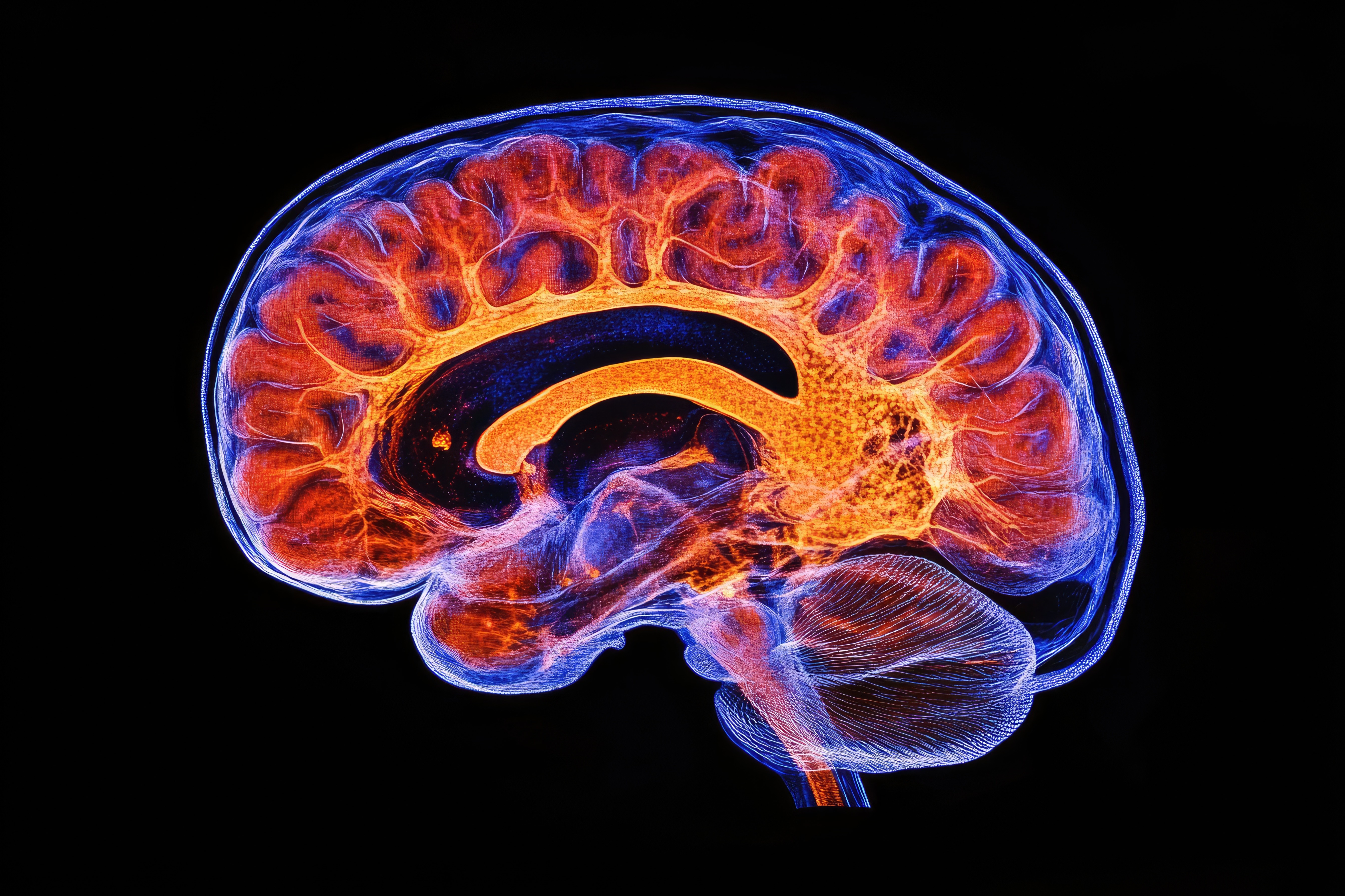
Brain MRI
Starting at 30 Minutes
A brain MRI (Magnetic Resonance Imaging) is a non-invasive imaging test that uses powerful magnets and radio waves to create detailed images of the brain and surrounding structures. Unlike X-rays or CT scans, it does not use radiation and is especially effective for visualizing soft tissues. A brain MRI is commonly used to evaluate symptoms such as headaches, seizures, dizziness, memory problems, or changes in vision and behavior. It helps detect a wide range of conditions, including brain tumors, stroke, multiple sclerosis, infections, aneurysms, brain injuries, and structural abnormalities. This advanced imaging technique provides critical information for accurate diagnosis, monitoring, and treatment planning.
09
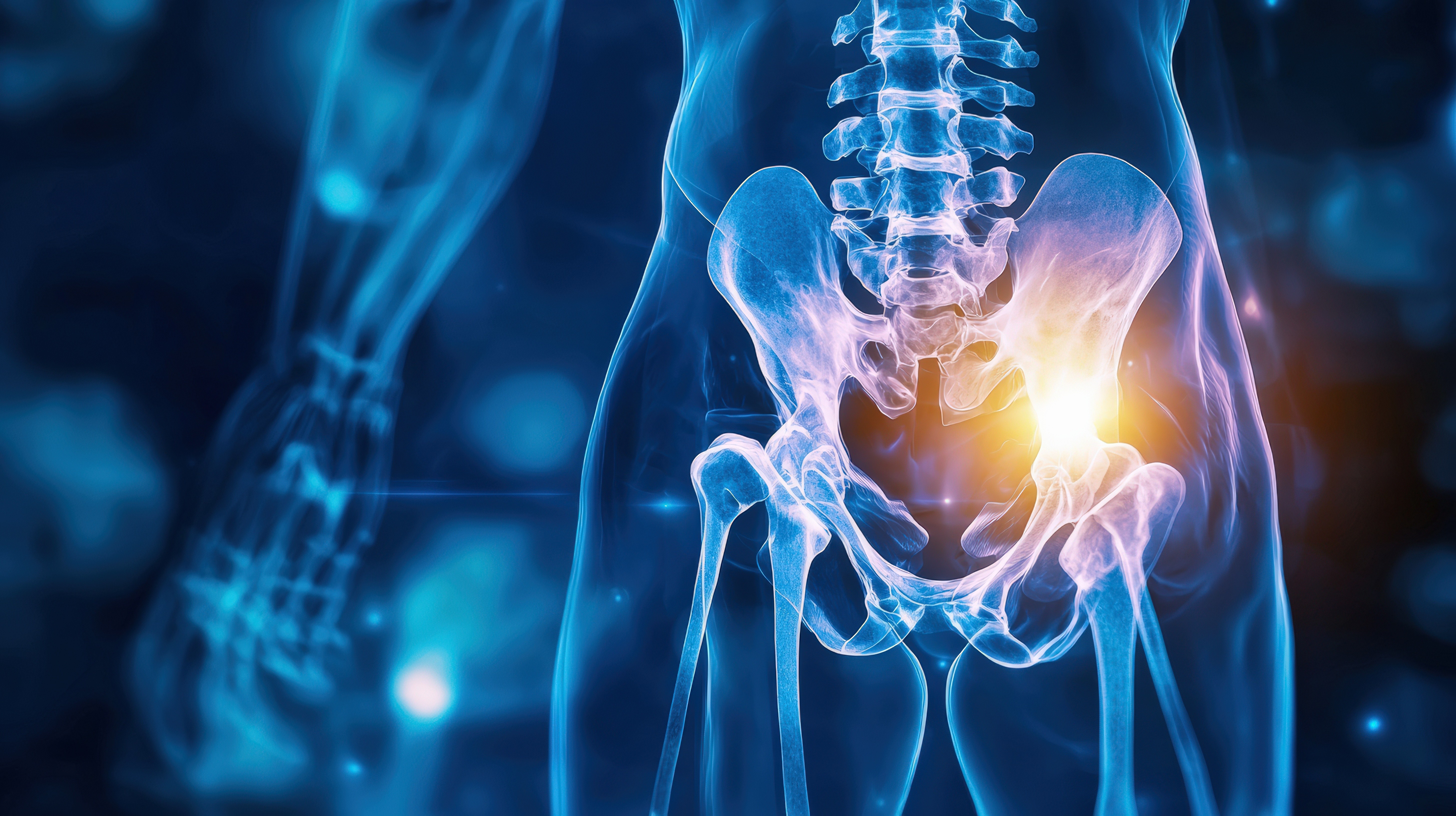
Hip MRI
Starting at 30 Minutes
A hip MRI (Magnetic Resonance Imaging of the hip) is a non-invasive diagnostic test that uses powerful magnets and radio waves to produce detailed images of the bones, cartilage, muscles, tendons, ligaments, and soft tissues surrounding the hip joint. It is commonly used to evaluate persistent hip pain, stiffness, swelling, or limited mobility—especially when X-rays or physical exams do not provide enough information. A hip MRI can detect conditions such as labral tears, hip impingement (FAI), bursitis, tendonitis, stress fractures, arthritis, and avascular necrosis. This advanced imaging tool helps physicians diagnose the root cause of symptoms and develop accurate, targeted treatment plans.
01
Cervical (Neck) MRI
A neck MRI is a powerful diagnostic tool used to evaluate the soft tissues, spinal structures, and blood vessels in the cervical spine. It is commonly recommended for individuals experiencing persistent neck pain, numbness or tingling in the arms, weakness, or after trauma such as whiplash. It also helps detect conditions like herniated discs, spinal stenosis, degenerative disc disease, tumors, and infections. Common findings may include disc bulges, nerve compression, arthritis, spinal cord abnormalities, or soft tissue masses—allowing for accurate diagnosis and effective treatment planning.
02
Thoracic (Mid-Back) MRI
A thoracic MRI is typically ordered to evaluate unexplained mid-back pain, neurological symptoms, or suspected spinal conditions affecting the upper spine. Doctors may recommend this scan for patients experiencing numbness, weakness, coordination issues, or symptoms unresponsive to other treatments. Common findings on a thoracic MRI include herniated or bulging discs, spinal stenosis, degenerative disc disease, spinal cord compression, and myelopathy. It can also detect more serious conditions like tumors, spinal cord cysts (syringomyelia), infections, fractures, or inflammatory diseases. This imaging is essential for accurately diagnosing the cause of symptoms and guiding effective treatment plans.
03
Lumbar (Low Back) MRI
A lumbar MRI is a valuable diagnostic tool used to evaluate lower back pain, sciatica, numbness, weakness, or symptoms following an injury. It’s commonly recommended for patients with chronic back pain, suspected disc herniations, degenerative disc disease, spinal stenosis, or nerve compression. This non-invasive scan provides detailed images of the spine’s bones, discs, nerves, and soft tissues, helping detect common issues such as bulging or herniated discs, arthritis, nerve impingement, spinal canal narrowing, spondylolisthesis, tumors, infections, or fractures. A lumbar MRI plays a key role in diagnosing the cause of symptoms and guiding effective treatment plans.
04
Shoulder MRI
A shoulder MRI is commonly ordered to evaluate pain, stiffness, weakness, or limited range of motion that doesn’t improve with rest or physical therapy. It’s especially useful after injuries, such as rotator cuff tears, labral tears, or dislocations, and helps diagnose chronic conditions like arthritis or tendonitis. This detailed imaging reveals common findings such as rotator cuff tears, labrum injuries, bursitis, tendon inflammation or degeneration, joint effusion, and arthritis. A shoulder MRI is a crucial tool for identifying soft tissue damage, guiding treatment decisions, and helping patients regain strength and mobility.
05
Knee MRI
A knee MRI is commonly performed to diagnose the cause of pain, swelling, instability, or limited movement in the knee joint. It is especially useful after injuries from sports, falls, or accidents, and helps identify conditions like ACL tears, meniscus injuries, ligament sprains, or cartilage damage. It also detects chronic issues such as osteoarthritis, tendonitis, bursitis, joint effusion, and Baker’s cysts. A knee MRI provides detailed images of the bones, ligaments, tendons, cartilage, and soft tissues, making it an essential tool for guiding treatment—whether that’s physical therapy, injections, or surgery.
06
Wrist MRI
A wrist MRI is a non-invasive imaging test that uses powerful magnets and radio waves to create detailed images of the bones, ligaments, tendons, cartilage, and soft tissues in the wrist. It is commonly used to evaluate unexplained wrist pain, injuries from falls, sports-related trauma, or symptoms like swelling, numbness, or weakness. A wrist MRI helps diagnose conditions such as ligament tears, TFCC injuries, tendonitis, carpal tunnel syndrome, arthritis, occult fractures, and ganglion cysts. This advanced imaging tool provides critical information for accurate diagnosis and effective treatment planning, especially when X-rays or physical exams are inconclusive.
07
Ankle MRI
An ankle MRI is a non-invasive imaging test that uses powerful magnets and radio waves to produce detailed images of the bones, ligaments, tendons, cartilage, and soft tissues in the ankle. It is commonly used to diagnose the cause of persistent pain, swelling, instability, or injury when X-rays or physical exams are inconclusive. An ankle MRI can detect ligament tears, Achilles tendon injuries, stress fractures, tendonitis, arthritis, and cartilage damage, providing critical information for accurate diagnosis and effective treatment planning.
08
Brain MRI
A brain MRI (Magnetic Resonance Imaging) is a non-invasive imaging test that uses powerful magnets and radio waves to create detailed images of the brain and surrounding structures. Unlike X-rays or CT scans, it does not use radiation and is especially effective for visualizing soft tissues. A brain MRI is commonly used to evaluate symptoms such as headaches, seizures, dizziness, memory problems, or changes in vision and behavior. It helps detect a wide range of conditions, including brain tumors, stroke, multiple sclerosis, infections, aneurysms, brain injuries, and structural abnormalities. This advanced imaging technique provides critical information for accurate diagnosis, monitoring, and treatment planning.
09
Hip MRI
A hip MRI (Magnetic Resonance Imaging of the hip) is a non-invasive diagnostic test that uses powerful magnets and radio waves to produce detailed images of the bones, cartilage, muscles, tendons, ligaments, and soft tissues surrounding the hip joint. It is commonly used to evaluate persistent hip pain, stiffness, swelling, or limited mobility—especially when X-rays or physical exams do not provide enough information. A hip MRI can detect conditions such as labral tears, hip impingement (FAI), bursitis, tendonitis, stress fractures, arthritis, and avascular necrosis. This advanced imaging tool helps physicians diagnose the root cause of symptoms and develop accurate, targeted treatment plans.